EchoBridgeMD Evidence Update #3: TEE Reveals Where We Are Actually Compressing During CPR


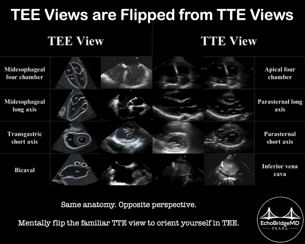
Transesophageal echocardiography (TEE) may allow clinicians to assess not only cardiac activity during arrest, but also whether chest compressions are actually being delivered over the optimal cardiac location.
In this prospective observational study, investigators performed point-of-care TEE immediately upon emergency department arrival in 18 patients with out-of-hospital cardiac arrest undergoing extracorporeal CPR with ongoing mechanical chest compressions. TEE successfully identified the area of maximal compression (AMC) in every patient.
Remarkably, chest compressions were appropriately aligned with the AMC in only 6 of 18 patients (33%). When compressions were aligned with the AMC, mean end-tidal CO₂ was substantially higher (33 vs 12.8 mmHg; p=0.004) and the odds of ROSC were significantly greater (OR 22; 95% CI 1.54–314; p=0.011). However, the study was small and observational, and no patient survived to hospital discharge, so whether TEE-guided optimization improves patient-centered outcomes was not fully answered.
At the bedside: Standard chest-compression landmarks do not guarantee optimal cardiac compression in every patient. TEE offers something unique during resuscitation: real-time visualization of what our compressions are actually doing to the heart.
Don’t just ask where you are compressing. Ask what you are compressing.
⠀
⠀
📖 Trainito G, Bernardo P, Bevilacqua S, et al. Usefulness of Point-of-Care Transesophageal Echocardiography in Cardiac Arrest to Identify the Area of Maximal Compression: A Prospective Observational Study. J Emerg Med. 2026.
⠀
⠀
⠀
🔗 Free subscription — receive future EchoBridgeMD educational content by email:

Comments